
Abstract
Obstructive sleep apnea syndrome (OSAS), a chronic condition characterized by collapse of the pharynx during sleep, has been increasingly recognized as a health issue of growing importance over the last decade. Recently emerging evidence suggests that there is a causal link between OSAS and hypertension, and hypertension represents an independent risk factor in OSAS patients. However, the pathophysiological basis for patients with OSAS having an increased risk for hypertension remains to be elucidated. The main acute physiological outcomes of OSAS are intermittent hypoxia, intrapleural pressure changes, and arousal from sleep, which might induce endothelial dysfunction, sympathetic activation, renin–angiotensin–aldosterone system activation, lipid metabolism dysfunction, and increased oxidative stress. This brief review focuses on the current understanding of the complex association between OSAS and hypertension.
Keywords: Approach, hypertension, obstructive sleep apnea syndrome, pathogenic mechanism
Introduction
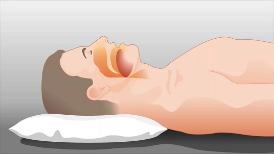
Sleep-related breathing disorders lead to concomitant alterations in the central nervous and cardiovascular systems, and such alterations often induce additional health issues. Sleep-disordered breathing represents a wide spectrum of sleep-related breathing abnormalities. Obstructive sleep apnea (OSA) is a condition in which there is repetitive partial or complete collapse of the pharynx during sleep. OSA associated with excessive daytime sleepiness is commonly called obstructive sleep apnea syndrome (OSAS). OSAS is the most common sleep-disordered breathing abnormality and often results in apnea or hypopnea, which can lead to snoring.
Our understanding of OSAS has evolved from initially regarding it as merely an annoying social situation to the recognition that OSAS may act through various mechanisms to increase cardiovascular risk and lead to increased morbidity and mortality. In particular, there is evidence that untreated OSAS may contribute to the pathophysiological mechanisms underlying the origin and/or development of hypertension, cardiac ischemia, myocardial infarction, congestive heart failure, and stroke. Of the different possible consequences of OSAS in patients, the most widely recognized may be the development of systemic hypertension. While many reviews have described the association between OSAS and hypertension, the diagnosis, prevalence, etiology, and new mechanisms linking OSAS to hypertension are outlined in this review.
Definition and diagnosis of OSAS

OSAS symptoms, including habitual and intermittent snoring, recurrent arousal during sleep, excessive daytime sleepiness, and witnessed apneas suggest the occurrence of OSAS. However, the gold standard diagnostic test for OSAS is the overnight in-laboratory polysomnography. Polysomnography uses multi-channel continuous recordings for electrocardiography, electromyography, electroencephalography, electro-oculography, nasal airflow, snoring sounds, blood oxygen saturation, thoracic and abdominal impedance belts for respiratory effort, and intra-esophageal pressure. The apnea-hypopnea index (AHI), defined as the average number of apneas and hypopneas per sleep hour, has always been used to evaluate OSAS severity. An apnea is defined as the complete airflow cessation for at least 10 seconds. Hypopnea may happen during sleep or awake states and fluctuate in severity of episodes of shallow breathing or an abnormally low respiratory rate. According to the American Academy of Sleep Medicine Task Force recommendations, OSAS is defined as an apnea-hypopnea index (AHI) >5, along with excessive daytime somnolence. Another diagnosis standard of OSAS is the number of apneas (where airflow stops completely) and obstructive hypopneas (> 50% reduction in respiratory flow or > 30% reduction linked to more than 3% desaturation and/or microarousals) lasting more than 10 seconds per hour. A threshold of 15 events per hour of recording is applied for OSAS diagnosis. OSAS is further subclassified into mild OSAS (AHI = 5–15), moderate OSAS (AHI = 16–30), and severe OSAS (AHI >30). A habitual snorer is a subject who always snores at night, while the AHI is <5.
Epidemiology of OSAS
The prevalence of OSAS varies among study populations, due to the use of different variables, and is influenced by the criteria used to define OSAS as well as by the population characteristics. The first large epidemiologic polysomnographic study of OSAS was done in 1995. Later, many studies reported the adult prevalence of OSAS in many different countries and among different ethnic groups. The overall estimated prevalence of OSAS is in the range of 3%–7% in adult men and 2%–5% in adult women, with certain subgroups of the population bearing higher risk. These subpopulations include overweight or obese people and middle-aged and older subjects. Notably, the results that there was no substantial difference in OSAS prevalence in North America, Europe, Australia, and Asia clearly suggest that OSAS is common not only in developed countries, but also in developing countries. In 2008, the American Heart Association and the American College of Cardiology distributed a joint scientific statement pointing out that 85% or more of humans with clinically significant OSAS have not been diagnosed. The referral populations of OSAS patients may represent only the tip of the iceberg of OSAS prevalence.
Evidence connecting hypertension to OSAS
OSAS is widespread in the middle-aged and older population, while hypertension is also highly prevalent among the middle-aged and older population. This raises the possibility of considerable co-morbidity between hypertension and OSAS. A lot of experimental and clinical evidence has indicated that the extent of the co-morbidity of hypertension and OSAS is actually substantially greater than expected.
In animal studies, direct evidence of the relationship between OSAS and hypertension has been well-established. Experimental OSAS resulted in acute transient increases in nighttime blood pressure (BP) and eventually produced sustained daytime hypertension. Acute normalization of blood pressure reduced sleep apneas in rats, even in the context of lifelong hypertension. Troncoso Brindeiro et al. developed a model of sleep apnea by exposing rats to chronic intermittent hypoxia (CIH) during sleep and found that this protocol increased blood pressure.
In humans, a large number of studies have sought to determine the presence and extent of a causal relationship between OSAS and hypertension, independent of frequently co-occurring and possibly confounding variables including age, body weight, and body mass index. The most convincing evidence for the support of a causal relationship between OSAS and hypertension has come from numerous epidemiological data collected in community populations. Early studies indicated that hypertension was found in about 50% of OSAS patients, while about 30% of hypertensive patients also have OSAS. The Wisconsin Sleep Cohort Study analyzed data on sleep-disordered breathing, blood pressure, habitus, and health history, at baseline and after 4 years of follow-up, in 709 Wisconsin state employees, using attended full polysomnography. Relative to the reference category of an apnea-hypopnea index of 0 events per hour at baseline, the odds ratios for the presence of hypertension was 2.03 (95% confidence intervals 1.29–3.17) in subjects with an apnea-hypopnea index of 5.0 to 14.9 events per hour, suggesting that the presence of hypertension was independent of known confounding factors and that sleep-disordered breathing is likely to be a risk factor for hypertension and subsequent cardiovascular morbidity in the general population. Moreover, the non-dippers exhibited a blunting of the sleep-related fall in blood pressure and an increased variability in blood pressure associated with sleep-disordered breathing. These results have been partly or completely confirmed by additional independent studies.
Some researchers have concluded that an increase in diastolic blood pressure might be the earliest hypertensive change associated with OSAS, evidenced by the fact that diastolic blood pressure was higher early in the course of OSAS, and diastolic and systolic-diastolic hypertension were the prominent types of hypertension observed both by clinical and ambulatory measurements. On the contrary, Sin et al. demonstrated that the systolic blood pressure was significantly higher in patients with OSA than in patients without OSA, indicating a high prevalence of systolic hypertension in patients with OSA. In addition, systemic hypertension was associated with a greater exacerbation of blood pressure variability in OSAS patients during sleep. The baroreflex sensitivity, an index of the cardiovascular control, was lower during wakefulness and rapid eye movement sleep in untreated OSAS patients than in normal subjects, and negatively correlated with the increase of blood pressure after apneas. Therefore, it seems that hypertension, increased blood pressure variability, and decreased baroreflex sensitivity may tightly correlate with OSAS and contribute to the increased cardiovascular risk of OSAS.
The similar risk factors in OSAS and hypertension
The pathophysiological pathways linking risk factors between OSAS and hypertension intersect with upper airway dilator muscle activity abnormalities, reduced arousal from sleep, reduced lung volume, and impaired ventilatory control stability. Community-based studies have identified some major risk factors for OSAS, including age, gender, and obesity. In parallel, these factors are also risk factors of hypertension. We summarize the similar risk factors between OSAS and essential hypertension in Figure 1, and discuss them below.
Convergence of epidemiological and biochemical variables in patients with OSAS and essential hypertension.
Age
The prevalence of OSAS increases with age. In older persons (≥ 65 years), the prevalence of OSAS is 2- to 3-fold higher than that in middle-aged individuals. However, the clinical and prognostic impact of OSAS in elderly patients appears lower than in young and middle-aged patients. Several studies have been conducted to identify the cause of the relationship between age and OSAS, but it seems that no consensus has been reached. Eikermann et al. reported that increasing age was correlated with both pharyngeal collapsibility and increase in pharyngeal resistance independent of body mass index (BMI) and gender during sleep, suggesting that the increased prevalence of OSAS in elderly individuals may mainly involve the dysfunction of muscles surrounding the parapharyngeal area. The prevalence of hypertension also increases with age. However, there is no satisfactory answer to the concurrent impact of age on OSAS and hypertension.
Obesity
During the past three or four decades, the United States has witnessed a dramatic increase in the number of overweight or obese individuals, which has become a public health issue. Epidemiologic studies from Europe and North America have clearly identified body weight as the strongest risk factor for OSAS. Respiratory function was certainly affected by obesity, and obesity could induce the collapse of upper airways during sleep. In addition, the frequency of respiratory events during sleep rises when body weight increases. An early report indicated that excess body weight was present in >60% of the sleep apnea patients. In the Wisconsin Cohort Study, a 10% weight gain predicted an approximately 32% increase in AHI and was associated with a 6-fold increased risk of OSAS, whereas a 10% weight loss predicted a 26% decrease in AHI, suggesting that the incidence of OSAS increases along with the increased incidence of obesity.
Longitudinal data from the Cleveland Family Study demonstrated that AHI was significantly associated with BMI and waist–hip ratio. Recently, it was demonstrated that among Americans aged 30–69 years, approximately 40% of adults with sleep-disordered breathing had BMIs ≥ 25, indicating obesity being a major risk factor for development of OSAS. Obesity is associated with anatomic changes that predispose to obstruction of the upper airway during sleep. These alterations may generate excess adiposity around the pharynx. Thus, the upper airway could be narrowed by the increases of adipose tissue in the neck and around the upper airway. Moreover, central obesity is associated with decrease of lung volume, which may cause an increase in pharyngeal collapsibility due to a loss of caudal traction of the upper airway.
However, in the Asian population, where visceral obesity is less prevalent, the prevalence of OSAS is not proportionately reduced. This phenomenon suggests that there may be a craniofacial effect interacting with body habitus. With obesity, the extra bulk of adipose tissue around the neck narrows the airway. A narrowed airway would increase the chances of airway collapse and closure during sleep. Mortimore and colleagues demonstrated that truncal and upper body obesity may be superior predictors of OSAS compared to body mass index, because of more fat deposition in the upper airway or pharynx. Flemons et al. also showed that increased neck circumference, which may be a marker for localized obesity, increased the risk of OSAS. Several imaging studies of patients with OSAS showed larger lateral parapharyngeal adipose tissues and pharyngeal walls in the neck and upper airway compared with non-obese controls. However, there have also been some inconsistent reports. Using magnetic resonance imaging, Schafer et al. reported that AHI significantly correlated to the amount of intra-abdominal fat, whereas there was no association between AHI and the size of parapharyngeal fat pads or subcutaneous fat of the neck region. The discrepancies may arise from different sets of populations and need to be further investigated.
This increasing prevalence of obesity and obesity-related hypertension has not only been described in the Western developed countries but also in developing countries such as China and India. Although obesity undoubtedly is a major risk factor of hypertension and OSAS, respectively, there is a lack of solid and clear clinical and experimental evidence for the in-depth mechanisms underlying the involvement of obesity between hypertension and OSAS. From a clinical viewpoint, nevertheless, a pragmatic approach to treating OSAS and preventing hypertension-related events would be desirable by treating obesity or reducing body weight.

Ethnicity
Most of the clinical population-based studies on OSAS prevalence were conducted in USA, Europe, and Australia. Recently, in Asian countries, including China, India, and Korea, several studies have been undertaken to characterize the burden of OSAS. The prevalence of OSAS in Asians is comparable to that documented in published reports of European and American populations. On the contrary, some studies suggest that the situation of OSAS may differ by race.
The overall prevalence of sleep-disordered breathing was approximately equal in Caucasians (30%) and African-Americans (32%), but the severity was higher in the African-American population. The odds ratio for severe sleep-disordered breathing was 2.55 for African-Americans compared to Caucasians, even after adjustment for BMI, sex, and age. In the Cleveland Family Study, Redline et al. found that African-Americans with sleep-disordered breathing were younger than Caucasians with sleep-disordered breathing. In addition, they showed that the association of body mass index with OSAS was stronger in Caucasians than in African-Americans. In the same subjects, brachycephaly was found to be associated with an increased AHI in Caucasians but not in African-Americans.
Similarly, a cross-sectional study of New Zealand Maori (Polynesian) and European (Caucasian) men showed that small reductions in mandibular prognathism and a wider bony nasal aperture represent major factors associated with OSAS in Polynesian men, whereas in the Caucasian group OSAS was associated with a larger neck circumference and a reduced retropalatal airway size. In addition, BMI was a stronger predictor of OSAS severity in Caucasian men compared with that in Polynesian men.
Ethnicity is associated with hypertension prevalence and is an important independent contributor to OSAS prevalence. The prevalence of OSAS in Japanese hypertensive patients was around 10%, which was one-third that of the Western hypertensive participants of the New York Sleep Heart Health Study. However, in the largest cross-sectional study involving a total of 6132 participants in China, the prevalence of hypertension in patients with OSAS was 56.2%, which is comparable to Western countries. The discrepancies among these results may need to be confirmed in other trials with larger number of patients.
Various categories of hypertension in OSAS
In normal subjects, blood pressure decreases during sleep by 10%–20% of the awake value and increases promptly on waking. An absence of this nocturnal dip in blood pressure correlates directly with the amount of deep sleep and inversely with indices of sleep fragmentation. Thus, there is great interest in the clinical observation and pathophysiological mechanisms underlying dipping and non-dipping patterns of ambulatory blood pressure profiles. In OSAS patients, various categories of hypertension were characterized.
Nocturnal hypertension
A nighttime fall of blood pressure (dipping) is normal. On the contrary, reduced nocturnal BP (non-dipping) or even higher nocturnal BP than daytime BP is an undoubted risk factor for hypertensive patients due to the end-organ damage and subsequent cardiovascular events. A blunted nocturnal BP dipping phenomenon is common in hypertensive patients. The differences between non-dipping and dipping may be related to the following: 1) alterations in the autonomic nervous function, 2) congestive heart failure, 3) chronic kidney disease, and 4) OSAS. Strikingly, the nocturnal BP profile described in studies of 24-hour blood pressure measurements in OSAS patients is similar to that in non-dipping hypertensive patients.
In the Wisconsin Sleep Cohort Study, Young and colleagues found a dose-response relationship between sleep-disordered breathing and 24-hour blood pressure, independent of known confounding factors. Baguet et al. found that 42% of apneic patients had clinical hypertension. In the study, 42% of the OSAS patients showed office hypertension, while 58% of subjects had daytime hypertension, and 76% of patients had nighttime hypertension.
Also, diastolic, systolic, and mean blood pressure values during sleep were significantly related to apnea-hypopnea index and age. Another study revealed that blood pressure night/day ratios in patients were associated with severity of OSAS. Moreover, a casual relationship between the increasing respiratory disturbance index and the average 24 hour systolic blood pressure was only observed in non-dipping individuals and hypertensive OSAS subjects. The repeated end-apneic arousal and/or hypoxic asphyxia and the subsequent sleep fragmentation contributed to nocturnal and diurnal elevation of BP. Furthermore, the hypertension is associated with a greater exacerbation of short-term variability during sleep in OSAS patients.
Moreover, the relationship between OSAS and nocturnal hypertension might differ between males and females. Interestingly, Portaluppi et al. studied 100 new cases of hypertension in men and found that non-dipping individuals exhibited a blunting of nocturnal blood pressure dipping phenomenon and an increased variability in BP associated with OSAS. This suggested that hypertensive non-dipping individuals had a high probability of coexisting sleep-disordered breathing. However, a significant relationship of nighttime/daytime blood pressure difference and AHI existed in men but not in women.
OSAS-related hypoxemia and hypercapnia, sleep fragmentation, increased sympathetic activity, chemoreflex activation, and nighttime blood pressure surges may be involved in the increased peripheral vascular tone (nocturnal hypertension) and subsequent cardiovascular events in OSAS patients (Figure 2). Cardiovascular events of relevance include increased incidence of platelet activation, obstructive cardiomyopathy, cardiac arrhythmias, myocardial ischemia or/and infarction, and sudden cardiac death. OSAS may also increase the incidence of hemorrhagic and ischemic stroke and subsequent neurological injury and cognitive impairment.
A schematic link between OSAS and nocturnal hypertension leading to cardiovascular events.
Resistant hypertension
OSAS is an important secondary cause of resistant hypertension and is particularly common in patients with resistant hypertension. Resistant hypertension, also called refractory hypertension, occurs where high blood pressure is not returned to within normal ranges with use of three drugs, including diuretics. A significant gender difference is typical, with OSAS being more prevalent and more severe in male than in female patients. Two cross-sectional studies demonstrate that the more severe the OSAS, the less likely blood pressure could be controlled despite increasing the number of antihypertensive agents. OSAS was extremely common in subjects with resistant hypertension, and a significant correlation between plasma aldosterone concentration and OSAS severity was observed in subjects with resistant hypertension but not in control subjects, suggesting that aldosterone excess may contribute to OSAS severity.
Masked hypertension
Masked hypertension is defined as a normal clinic blood pressure and elevated out-of-clinic blood pressure, assessed using self-monitoring of blood pressure by the patients at home. Masked hypertension includes stress-induced hypertension, morning hypertension, and nocturnal hypertension, which is defined as a sleeping blood pressure >120/70 mmHg. Recently, Baguet et al. showed a large incidence of masked hypertension, as determined by ambulatory BP monitoring (ABPM), in patients with OSAS who were considered normotensive. The authors concluded that masked hypertension is frequently underestimated in OSAS and is nearly always present when clinic blood pressure is >125/83 mmHg.
A recent study revealed that among apparently normotensive male OSAS patients, masked hypertension is present in one-third of patients and there is a progressive impairment of arterial stiffness in OSAS patients with masked hypertension, indicating that the diagnosis of masked hypertension may be underestimated in OSAS patients and that OSAS has an association with arterial stiffness independent of masked hypertension. Moreover, obesity may be involved in the relationship between OSAS and masked hypertension. Although common etiologies of masked hypertension include stressful living situations, sleep apnea resulting from obesity is also a possible etiology. However, the relationship between masked hypertension and OSAS needs more study.
Pulmonary hypertension
The first report regarding OSAS and pulmonary hypertension was in 1988. No significant correlation existed between pulmonary arterial pressure and the apnea index. However, while intravascular pulmonary arterial pressure decreased during apneas and increased at the resumption of breathing, transmural pulmonary arterial pressure showed a progressive increase during apneas that decreased once ventilation had been resumed, suggesting pulmonary arterial pressure might be related with OSAS. The pulmonary artery hypertension was linked to the presence of an obstructive ventilatory pattern, hypoxemia, and hypercapnia, while the severity of OSAS plays only a minor role. In a study that recruited 92 consecutive patients, OSAS was found to be an important independent risk factor for pulmonary hypertension.
Mechanism underlying the link between OSAS and hypertension
The main acute physiological consequences of OSAS are intermittent hypoxia, intrapleural pressure changes, and arousals, which might induce endothelial dysfunction, sympathetic activation, lipid metabolism dysfunction, increased oxidative stress, etc. (Figure 1). All of these consequences could increase the artery tone and arterial stiffness, and thereby increase the risk of systemic hypertension and further cardiovascular diseases such as stroke and atherosclerosis.
Intermittent hypoxia
Intermittent hypoxia, particularly if associated with episodes of intermittent reoxygenation, is one of the most important features in OSAS. Intermittent hypoxia causes remarkable blood oxygen desaturation, commonly within a cycle time of less than 1 min. To a certain degree, the intermittent hypoxia induced a repetitive hypoxia/reoxygenation cycle in OSAS, resembling ischemia-reperfusion injury, by promoting the production of reactive oxygen species (ROS), activating systemic inflammation, and ultimately impairing endothelial function. Apart from the induction of systemic inflammation and oxidative stress, intermittent hypoxia increased the plasma level of vasoconstrictive endothelin-1. Additionally, intermittent hypoxia has been proposed to enhance peripheral chemoreceptors and the sympathetic nervous system activity. Moreover, hypoxia-induced cellular responses, such as apoptosis and autophagy, have to be considered.
Endothelial dysfunction
Endothelial dysfunction is a systemic pathological state of the vascular endothelium and can be broadly defined as an imbalance between vasorelaxation and vasoconstriction substances produced by the endothelium. Endothelial dysfunction is an early marker of vascular damage that precedes clinically overt vascular disease and might be an important promoter of cardiovascular events in patients with OSAS. The strong association between OSAS and cardiovascular diseases may be due to significant endothelial dysfunction in OSAS patients and therefore is accumulating evidence for the association of OSAS and the impaired endothelial function/reduced endothelial repair capacity.
A previous study showed that even mild OSAS was associated with reduced endothelium-dependent vasodilatation. Greater impairment of endothelial function was undoubtedly associated with more severe OSAS. Studies on hypertensive patients with OSAS revealed that there indeed is an association between OSAS and endothelial dysfunction. Furthermore, in normotensive patients with OSAS, endothelium-dependent vasodilatation, measured by forearm blood flow, was impaired.
Nitric oxide (NO), the most important vasodilatory molecule synthesized by the endothelium, decreased in patients with OSAS. In addition, the level and activity of endothelial progenitor cells, a population of rare cells that circulate in the blood with the ability to differentiate into endothelial cells, decreased in patients with OSAS. Many above-mentioned signaling pathway proteins, including tumor necrosis factor, interleukins IL-1, IL-6, and IL-8, nuclear factor κB (NF-κB), the renin–angiotensin system, and hypoxia-inducible factor-1 (HIF-1) participate in the molecular mechanisms underlying the OSAS-induced endothelial dysfunction. These signaling proteins will be discussed in the next section.
Inflammation and oxidative stress
The evidence that OSAS is associated with increased oxidative stress and inflammation is based on findings from both animals and humans. Promoting inflammation and oxidative stress is a major cause of endothelial dysfunction by decreasing NO availability and reducing endothelial vasodilatation capacity. Cells exposed to intermittent hypoxia demonstrated selective activation of the pro-inflammatory transcription factor NF-κB, whereas the adaptive regulator HIF-1 was not activated. In addition, elevated NF-κB activity induced by chronic intermittent hypoxia was accompanied by increased expression of inducible nitric oxide synthase (iNOS) level, a putative and important NF-κB-dependent inflammatory and oxidative protein.
Moreover, NF-κB could further stimulate the production of pro-inflammatory mediators such as IL-8 and intercellular adhesion molecule 1 (ICAM-1). TNF-alpha levels are independently associated with excessive daytime sleepiness, and IL-8 has shown elevated levels in patients with OSAS compared with controls. A recent study raised the possibility that the obese OSAS patients might have elevated TNF-alpha levels compared to BMI-matched controls. These factors may contribute to the hypertension in OSAS. C-reactive protein (CRP) plays an important role in hypertension and is correlated with OSAS. Shamsuzzaman et al. reported that plasma CRP levels were significantly higher in patients with OSAS than in controls, suggesting that the severity of OSAS is proportional to the CRP level, which was confirmed later by numerous cross-sectional, case-control, and non-randomized interventional studies. Apart from these systemic investigations, cellular studies on lung might also provide some association between inflammation and OSAS.
Management and treatment of OSAS
Non-drug therapy
Weight loss is an effective treatment of obesity in OSAS patients. Weight loss improves significantly sleep apnea and has favorable effects on blood pressure and baroreflex sensitivity. The improvement of obstructive sleep apnea after weight loss might be related to improvement in pharyngeal and glottic function. Avoidance of alcohol is to be recommended as deterioration of OSAS occurs by alcohol abuse.
Continuous positive airway pressure (CPAP) therapy
The most common and highly efficient therapeutic procedure of eliminating airway obstruction is continuous positive airway pressure (CPAP) therapy, a form of treatment first described in 1981 by Sullivan et al. This therapy exerts a blood pressure-lowering effect, reduces nocturnal sympathetic nerve traffic, blunts blood pressure surges, decreases nocturnal blood pressure surge, and improves cardiovascular prognosis in many OSAS patients. The benefit was larger in patients with more severe sleep apnea than in those who had less severe apnea, but was independent of the baseline blood pressure.
However, there are also some studies reporting negative effects. Lack of concordance may be due to heterogeneity of selected subjects and study design. Thus, three meta-analyses were employed to summarize findings of intervention studies in CPAP treatment. Bazzano et al. reported that mean net change in systolic blood pressure for those treated with CPAP application of 2–24 weeks (818 patients) compared with controls was –2.46 mmHg (P < 0.05). Another meta-analysis included 12 trials (572 patients) and reported a significant –1.69 mmHg decrease in mean BP, with similar reductions in SBP and DBP (P < 0.05).
On the contrary, Alajmi et al. reported that the effects of CPAP on BP in OSAS patients (572 patients) were modest and not statistically significant (P = 0.23). The authors considered that in unselected patients with sleep apnea, CPAP may have modest effects on BP, whereas they could not exclude the possibility that certain subgroups of patients may have more robust responses to CPAP. Obviously, although CPAP has been the first-line therapeutic strategy for OSAS, the beneficial effect of CPAP remains an open question.
Conclusions
Over the last decade, there has been increased interest in OSAS-related research and increased understanding of the OSAS-related cardiovascular complications. Because OSAS is associated with hypertension and hypertension associated end-stage organ diseases such as stroke, coronary heart disease, and arrhythmia, the employment of CPAP is highly encouraged as CPAP therapy seems to assist blood pressure control at least in those with severe apnea, resistant hypertension, and daytime sleepiness. This field continues to be updated monthly. A number of key questions, however, remains to be resolved. More clinical research is warranted to characterize more fully the underlying mechanisms and to develop practicable strategies for OSAS treatment.
Name | Position | Office | Age |
 | 25 Ways To Stay Calm In Stressful Situations | ||
 | 17 Ways To Get Happy When You Are Sad | ||
 | 20 Ways About How to Calm Yourself During an Anxiety Attack | ||
 | 13 Ways To Calm Yourself Down When You’re Nervous |